
- News
Immunity (Impact Factor: 30.6): Shanghai Jiao Tong University Team Led by Wang Honglin Uncovers a Novel Neuroimmune Mechanism and Therapeutic Strategy for Vitiligo
Vitiligo is a chronic autoimmune skin disease characterized by localized or widespread white patches caused by the destruction of melanocytes, the cells responsible for skin pigmentation. Beyond its visible effects, vitiligo can profoundly affect patients’ education, social interactions, careers, and personal relationships. The condition may also lead to low self-esteem, depression, severe psychological distress, and, in some cases, suicidal thoughts or behavior. Vitiligo affects approximately 0.5%–2% of the global population [1]. In China, the number of people living with the condition reached 22.83 million in 2021 and has continued to rise. The current estimate is approximately 30 million patients nationwide. More than half develop vitiligo before the age of 20, and the disease can persist throughout life, making it difficult to cure.
The causes of vitiligo are complex and incompletely understood, and effective targeted therapies remain scarce. Historically, treatment has relied heavily on off-label drug use (OLDU) and nonspecific immunosuppressants, including glucocorticoids and calcineurin inhibitors. These treatments have limited efficacy and safety concerns, particularly with long-term use, which may cause skin atrophy and increase the risk of infection. As a result, they often fall short of meeting patients’ long-term treatment needs. The development of new, safe, and effective therapies is therefore an urgent global priority. Although the approval of ruxolitinib cream marked a major advance, substantial unmet clinical needs remain. In particular, ruxolitinib has not yet been approved for use in China, leaving approximately 30 million Chinese patients without access to an approved targeted therapy for vitiligo.
On June 16, 2025, the team led by Professor Wang Honglin at Shanghai General Hospital, Shanghai Jiao Tong University School of Medicine, published an online research article in Immunity entitled “Nociceptor-derived CGRP enhances dermal type I conventional dendritic cell function to drive autoreactive CD8+ T cell responses in vitiligo.”
The study identifies a previously unrecognized disease-driving pathway in vitiligo: the sensory neuron–CGRP–cDC1–CD8+ T cell axis. By blocking this neuroimmune pathway with the CGRP receptor antagonist rimegepant, the researchers significantly inhibited disease progression in a mouse model of vitiligo. A clinical trial involving 57 patients also demonstrated promising therapeutic effects, offering a new strategy for vitiligo treatment.
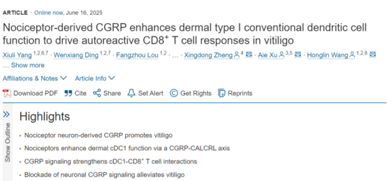
Image source: Immunity
https://www.cell.com/immunity/fulltext/S1074-7613(25)00239-0
Study Design and Methods
The researchers generated several genetically engineered mouse models, including Nav1.8-tdTomato sensory neuron reporter mice, Nav1.8-tdTomato-DTR mice for conditional sensory neuron ablation, and mice with cDC1-specific deletion of the CGRP receptor gene (Xcr1-Cre; Calcrl^fl/fl^, referred to as cKO mice).
By integrating single-cell sequencing, spatial transcriptomics, whole-mount tissue-clearing imaging of the skin, nearly 10 genetically engineered mouse models, and an investigator-initiated trial (IIT), the team established a comprehensive research framework and uncovered the neuroimmune mechanism underlying vitiligo.
Key Technical Approaches
1. Integrated single-cell and spatial transcriptomic analyses
2. Whole-mount immunofluorescence staining of clinical skin samples
3. Development of a mouse model that closely recapitulates human vitiligo
4. Mechanistic investigations using nearly 10 genetically engineered mouse models
5. Validation through an investigator-initiated clinical trial
Key Findings
1. Identifying a potential interaction between CGRP+ sensory nerves and dermal cDC1s in affected skin in Vitiligo Lesions
To address the lack of targeted treatments for vitiligo, the team led by Professor Wang Honglin at Shanghai General Hospital collaborated with Professor Xu Ai’e’s team at Hangzhou Third People’s Hospital.
The researchers first performed integrated single-cell and spatial transcriptomic analyses of healthy and vitiligo-affected skin. They identified interactions between type 1 conventional dendritic cells (cDC1s) and CD8+ T cells in vitiligo lesions. Notably, dermal cDC1s specifically expressed high levels of the CGRP receptor components CALCRL and RAMP1, suggesting that these immune cells may be regulated by CGRP signaling.
The skin is a complex, highly coordinated organ containing diverse nerve fibers, immune cells, and resident tissue cells. Cutaneous sensory nerves originate from neuronal cell bodies in the dorsal root ganglia (DRG) of the spinal cord. Among them, Nav1.8+/TRPV1+ sensory neurons can extend into the epidermis and release the neuropeptide calcitonin gene-related peptide (CGRP), which regulates a range of cutaneous immune responses and plays an important role in neuroimmune communication.
In previous studies, Professor Wang’s team demonstrated that sensory neurons promote psoriasis through CGRP signaling and that targeting sensory nerves with lidocaine can effectively treat psoriasis (Journal of Investigative Dermatology, 2022). The team subsequently identified a TNC+ dermal fibroblast subpopulation that promotes abnormal nerve sprouting and neuroimmune interactions in psoriasis, offering new insights into the remodeling of the local immune microenvironment in inflammatory skin diseases (Nature Communications, 2023).
Building on the finding that dermal cDC1s in vitiligo express high levels of CGRP receptors, the researchers hypothesized that CGRP-producing sensory neurons might also contribute to vitiligo development. Using whole-mount skin clearing and imaging, they found that both the density of CGRP+ sensory nerves and the number of dermal cDC1s were significantly increased in vitiligo lesions compared with healthy skin. The two cell populations were also located in close spatial proximity, suggesting a potential interaction between CGRP+ sensory nerves and dermal cDC1s in affected skin (Figure 1).
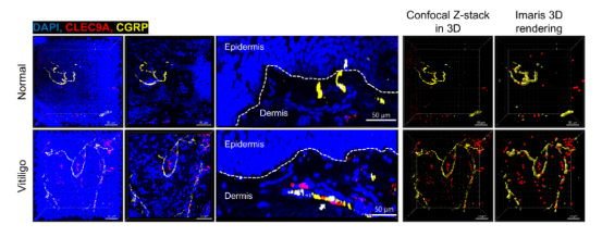
Figure 1. CGRP+ sensory nerve density and dermal cDC1 numbers are elevated in vitiligo lesions, with the two populations located in close proximity [1].
2. Sensory Neurons and CGRP Drive Vitiligo Lesions Progression in a Mouse Model
The team established a mouse model of vitiligo using B16F10 melanoma cells with CD4+ T-cell depletion, designed to closely recapitulate key features of human disease.
Using Nav1.8-tdTomato sensory neuron reporter mice, the researchers found extensive colocalization between Nav1.8+ nerves and CGRP+ nerves, indicating that Nav1.8+ sensory neurons are a major source of CGRP in the skin. As in human vitiligo lesions, the model mice also exhibited increased CGRP+ nerve density and increased numbers of dermal cDC1s.
The researchers then selectively ablated sensory neurons using Nav1.8-tdTomato-DTR mice or resiniferatoxin (RTX). In both cases, sensory nerve ablation significantly inhibited disease progression in the vitiligo model. To determine whether neuron-derived CGRP was directly involved, the team administered exogenous CGRP to mice after sensory nerve ablation. This restored the vitiligo-like disease phenotype to a substantial degree.
Together, these findings demonstrate that sensory neurons and the CGRP they release contribute to the development and progression of vitiligo in the mouse model.
3. Dermal cDC1s Are Essential for Activating Autoreactive CD8+ T Cells
The researchers observed significantly increased numbers of dermal cDC1s in both patients with vitiligo and the mouse model. To investigate their role in disease, the team generated Xcr1-tdTomato reporter mice to label cDC1s and examine their morphology.
Following vitiligo induction, dermal cDC1s changed from a rounded shape to a more dendritic morphology, consistent with an activated state. These cells were also closely associated with CGRP+ nerve fibers, further supporting a potential neuroimmune interaction (Figure 2).
Experiments involving the depletion or expansion of cDC1s in vivo provided additional evidence that dermal cDC1s are essential for initiating autoreactive CD8+ T-cell responses and play a critical role in vitiligo development and progression in mice.
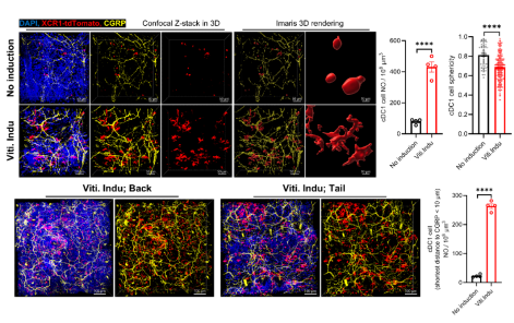
Figure 2. Dermal cDC1s are activated in vitiligo-model mice and closely interact with CGRP+ sensory nerves [1].
4. CGRP Signaling Regulates cDC1 Function and Amplifies Autoimmune Responses
To establish whether sensory neuron-derived CGRP directly regulates dermal cDC1s, the team used Calcrl^fl/fl^ mice to generate cDC1-specific CGRP receptor knockout mice (Xcr1-Cre; Calcrl^fl/fl^, cKO).
Disease severity was significantly reduced in cKO mice compared with control animals. This finding indicates that sensory neurons regulate dermal cDC1 function through the CGRP–CALCRL signaling pathway, thereby influencing vitiligo progression (Figure 3).
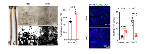
Figure 3. cDC1-specific deletion of the CGRP receptor alleviates disease in the mouse model of vitiligo [1].
The team next established several in vitro co-culture systems to investigate the effects of sensory neurons and CGRP on cDC1 function. Both neuronal co-culture and direct CGRP treatment enhanced the antigen-presenting capacity of cDC1s and significantly increased the ability of CD8+ T cells to produce interferon-gamma (IFN-γ).
Transcriptomic changes in cDC1s exposed to sensory neurons closely resembled those induced by CGRP treatment. The upregulated genes were significantly enriched in pathways associated with T-cell-mediated cytotoxicity and IFN-γ responses. When cDC1s from cKO mice were used, the effects of neuronal co-culture were markedly attenuated, indicating that sensory neurons regulate cDC1 function primarily through CGRP signaling.
Immunofluorescence staining of skin from vitiligo-model mice further revealed excessive cDC1 activation and close associations between cDC1s and CGRP+ sensory nerves. Large numbers of clonally expanded CD8+ T cells were found around cDC1s, with some migrating toward the dermal–epidermal junction, where melanocytes reside.
Integrating these in vivo and in vitro findings, the team identified a previously unrecognized disease-driving pathway in vitiligo: the sensory neuron–CGRP–cDC1–CD8+ T cell axis (Figure 4).

Figure 4. A novel neuroimmune mechanism in vitiligo: the sensory neuron–CGRP–cDC1–CD8+ T cell axis [1].
5. Rimegepant Emerges as a Potential New Treatment Strategy
A central motivation for the study was the urgent need for targeted vitiligo therapies. Guided by their discovery of the neuroimmune pathway, the researchers investigated whether blocking the sensory neuron–CGRP–cDC1–CD8+ T-cell axis could effectively treat the disease.
CGRP-pathway inhibitors are already used clinically to treat migraine. Approved therapies include four monoclonal antibodies targeting CGRP or its receptor and four small-molecule CGRP receptor antagonists. Given the potential need for topical and long-term treatment in vitiligo, the team focused on small-molecule compounds that could potentially be developed into topical formulations.
Rimegepant, an orally disintegrating tablet approved in China in 2024 for migraine treatment, was selected as the lead candidate.
The team first evaluated rimegepant in the mouse model of vitiligo. Both rimegepant and ruxolitinib significantly improved disease manifestations, with no significant difference in their overall effects. However, flow cytometry showed that rimegepant inhibited the expansion of dermal cDC1s more strongly than ruxolitinib, suggesting a potential advantage in modulating the disease-associated immune response.
The researchers subsequently developed a topical ointment formulation of rimegepant and initiated a single-blind clinical trial involving 57 patients. Participants received treatment for 6–12 weeks and were assigned to three study groups:
* Group A: Treatment with either a placebo or 0.1% rimegepant ointment.
* Group B: Treatment with either a placebo or 0.1% rimegepant ointment, combined with once-weekly narrowband ultraviolet B (NB-UVB) phototherapy.
* Group C: Two adjacent vitiligo patches on the same patient were selected for comparison. One was treated with placebo ointment and the other with 0.1% rimegepant ointment, with both sites receiving phototherapy.
Across all three groups, rimegepant ointment promoted repigmentation and significantly increased the rate of repigmentation in vitiligo lesions.
Taken together, the animal experiments and clinical findings demonstrate the therapeutic potential of CGRP receptor antagonism in both experimental and human vitiligo (Figure 5).
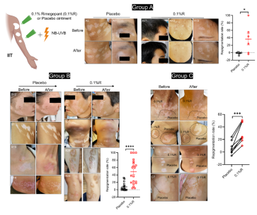
Figure 5. Topical 0.1% rimegepant ointment significantly improves vitiligo manifestations in patients [1].
Conclusions and Clinical Significance
This study demonstrates that sensory neurons in vitiligo lesions release CGRP, which regulates the functional state of dermal cDC1s. CGRP signaling enhances their antigen-presenting capacity and expression of costimulatory molecules, thereby initiating and amplifying autoreactive CD8+ T-cell responses against melanocytes. The resulting immune attack progressively destroys epidermal melanocytes, leading to the characteristic depigmented patches of vitiligo.
Blocking the pathogenic CGRP–CALCRL signaling axis with the CGRP receptor antagonist rimegepant significantly suppresses cDC1 expansion and cytotoxic CD8+ T-cell responses. This may help establish a more immunosuppressive local environment that supports melanocyte regeneration and migration stimulated by phototherapy.
Importantly, the research directly addresses the shortage of targeted treatments for vitiligo. By identifying the sensory neuron–CGRP–cDC1–CD8+ T-cell axis, the team has established a new mechanistic framework and potential therapeutic target for future drug development. The team’s reformulated 0.1% topical rimegepant ointment also showed promising results in a clinical trial, completing a translational research cycle that connects clinical needs, basic mechanistic discoveries, and clinical validation.
These findings open a promising avenue for developing neuroimmune-targeted treatments for vitiligo. Further studies will be needed to establish the formulation’s efficacy and safety, determine optimal dosing and treatment duration, and assess its potential for broader clinical use.
Reference:
[1]Yang et al., Nociceptor-derived CGRP enhances dermal type I conventional dendritic cell function to drive autoreactive CD8+ T cell responses in vitiligo, Immunity (2025), https://doi.org/10.1016/j.immuni.2025.05.018
Source:https://mp.weixin.qq.com/s/gpH9WlcCg1ngM7Y06DMpQQ?scene=1&click_id=844145141