- News
Hepatobiliary Surgery Team from Shanghai General Hospital Accurately Removed a Giant Hepatic Hemangioma from a Pregnant Patient
Recently, the Hepatobiliary Surgery team of our hospital successfully removed a giant hepatic hemangioma weighing 2.63 kilograms from a special pregnant woman's body. The tumor grows rapidly during the patient's pregnancy, with a maximum diameter of nearly 30 centimeters, completely surrounding the core blood vessels of the liver and causing multiple organs to be compressed. As a result, continuing the pregnancy will put the patient at extremely high risk of life. Faced with multiple challenges such as huge tumors, complex anatomical structures, moderate anemia, and coagulation disorders in patients, the team led by Director Xu Junming of the Hepatobiliary Surgery Department at the General Surgery Center, in collaboration with gynecology, radiology, anesthesia, and transfusion departments, successfully completed a high difficulty resection of the right lobe and caudate lobe which lasted for 3 hours to resolve the crisis. At present, the patient has recovered and has been discharged from the hospital.
About a week ago, a young woman came to our hospital for treatment due to early pregnancy. After receiving the consultation, Prof. Wu Hao, Deputy Director of Obstetrics, found that her abdominal circumference was much larger than the actual gestational age. After further examination, a huge solid mass was found in the patient's abdominal cavity, which is suspected to be a giant hemangioma of the liver. Given the enormous size of the tumor which has already caused compression on adjacent organs, continuing the pregnancy would pose a significant risk to the lives of both the pregnant woman and the fetus. The obstetrics department immediately requested a consultation with the hepatobiliary surgery department.
After a detailed discussion, Prof. Xu Junming's team from hepatobiliary surgery department assessed that the tumor had a maximum diameter of nearly 30cm and completely surrounded the first and second porta hepatis, causing compression and deformation of organs such as the kidneys and gastrointestinal tract. This surgery faces four major risks: firstly, the long-term compression of the huge tumor leads to rotation of the liver axis and deformation of the hepatic portal duct structure, making preoperative CT vascular imaging reconstruction used to guide the surgery exceptionally difficult; Secondly, radiological imaging shows congenital variations in the hepatic artery, shallow course of hepatic veins, and complex anatomical structures; Thirdly, the tumor occupies the majority of the abdominal cavity, resulting in extremely narrow operating space and difficulty in distinguishing local structures. A slight mistake during surgery may cause tumor rupture and massive bleeding; Fourthly, the patient still had moderate anemia and hyperfibrinolysis before surgery, further increasing the risk of surgical tolerance.
Faced with such complex condition, the surgical team developed a meticulous "three-step" treatment strategy after sufficient communication with the family. The first step is to terminate the pregnancy as soon as possible; The second step is to timely perform the intervention treatment for liver hemangioma after the end of the perinatal period to promote tumor shrinkage and healthy left liver hyperplasia; The third step is to perform the radical surgical resection. After completing the first two steps as scheduled, the intervention therapy achieved significant results, reducing the tumor length from 28cm to 19.5cm, creating favorable conditions for the final surgery.
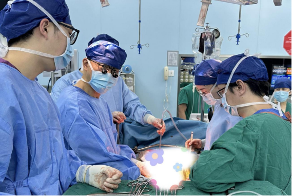
On the day of the surgery, Prof. Xu Junming's team, with the support of the anesthesia and transfusion departments, accurately controlled the blood vessels entering and leaving the liver, successfully achieving anatomical right hemihepatectomy and caudate lobe resection. The entire surgery lasted for 3 hours, and the patient's hemoglobin level remained stable during the operation without the need for additional blood transfusion. Finally, the tumor with a net weight of 2.63 kilograms was successfully removed. Under the guidance of the accelerated rehabilitation concept, the patient was able to get out of bed and move around on the second day after surgery, and successfully recovered and was discharged on the sixth day after surgery.