- News
Intelligent Exploration of Tumor Radiotherapy: A Series of Innovative Achievements by Professor Liu Yong's Team in the Department of Radiology at Shanghai General Hospital.
https://mp.weixin.qq.com/s/01g_Cf2Gqp0-L9U0cIX20A
Radiation therapy is one of the primary methods for cancer treatment, involving multiple disciplines such as clinical oncology, medical physics, radiodosimetry, radiological technology, and radiobiology. It is characterized by a distinct integration of medicine and engineering, with profound theoretical underpinnings, intricate processes, and complex techniques. The integration of artificial intelligence (AI) presents new opportunities to optimize radiation therapy.
As a frontline clinician with extensive interdisciplinary research experience, Professor Liu Yong from the Radiation Oncology Department at Shanghai General Hospital is well-versed in the challenges encountered in clinical radiation oncology. In response to these complex issues, Professor Liu Yong and his team have dedicated years to exploring the application of AI in cancer radiation therapy, investigating its value from various perspectives. Their research has yielded a series of significant findings, addressing three core clinical challenges, with papers published in internationally renowned academic journals, including Nature Machine Intelligence and Radiotherapy and Oncology.
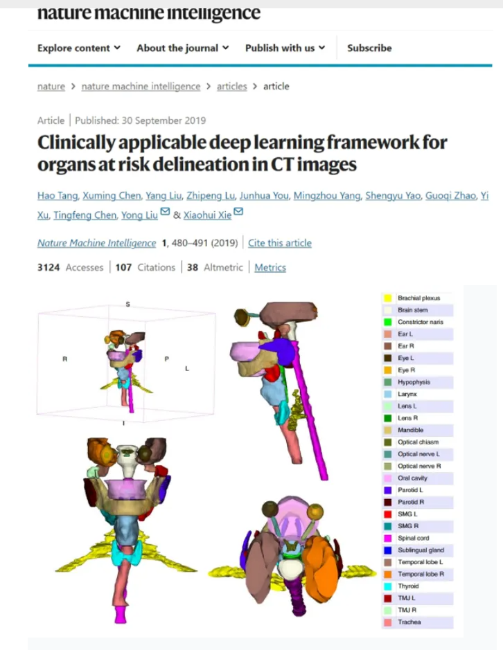
Outlining normal tissues on radiotherapy planning CT images is an essential step in radiotherapy workflows. In previous routine practices, manual outlining of a patient's organs typically took more than 30 minutes, which was not only time-consuming and labor-intensive but also prone to individual variability. To address this issue and improve efficiency and accuracy, the team at Shanghai General Hospital collaborated with Professor Xie Xiaohui’s team from the University of California to develop AI algorithms for automated outlining of normal tissues.
Over the course of three years, the team successfully completed research on automatic delineation of 28 normal tissues in the head and neck region. The study introduced a modified U-Net architecture, which further improved the accuracy of automated outlining and significantly reduced the time required for both model training and tissue delineation. This achievement reduced the original manual outlining time from over 30 minutes to under one minute, with an accuracy comparable to that of clinical experts, fully meeting clinical application standards.
The results were published in 2019 in Nature Machine Intelligence, an AI-focused journal. Notably, this was the first radiotherapy research published in this journal since its inception (Clinically applicable deep learning framework for organs at risk delineation in CT images. Nat Mach Intell, 2019, 1, 480–491).
Radiation therapy can be applied not only to the head and neck but also to various organs throughout the body. Therefore, the automatic delineation of normal tissues must address the needs of multiple organs across the body. However, the delineation of organs in different regions poses challenges due to variations in algorithmic models. Identifying the optimal delineation automatically is essential for improving both efficiency and precision.
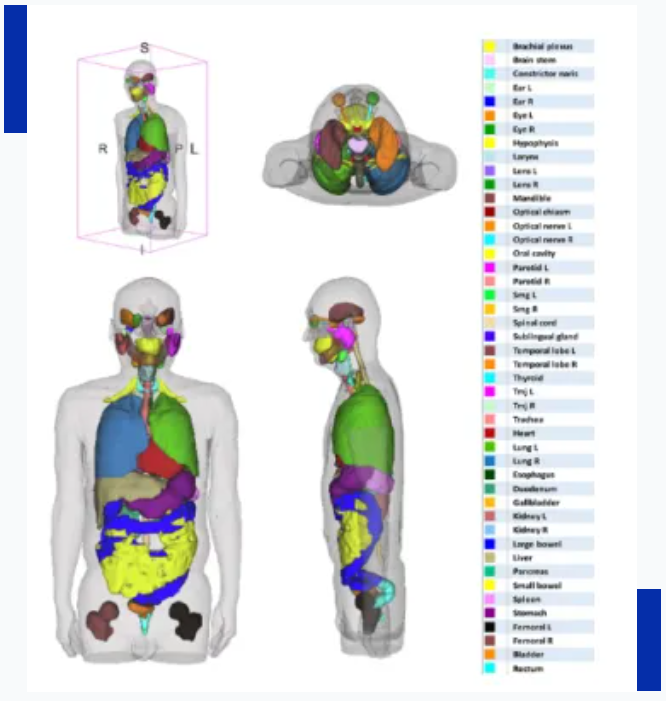
Based on this clinical need, the team expanded the scope of automatic delineation from head and neck tissues to the entire body, completing a study involving the automatic delineation of 50 normal tissues and organs, covering the head and neck, thoracic, and abdominopelvic regions. The study proposed a novel whole-body automatic delineation framework called WBNet, which is not restricted by image location. It can automatically identify anatomical regions in CT images and match appropriate algorithms to perform delineation based on the anatomical characteristics of different areas.
This achievement addresses all common challenges in the automatic delineation of normal tissues during radiotherapy. As a new in-house technology at Shanghai General Hospital, it has been routinely applied in the Department of Radiotherapy’s daily operations, assisting doctors in delineating normal tissues for more than 3,000 patients to date. This system has saved considerable time by reducing repetitive tasks and has significantly improved work efficiency.
The results were published in 2021 in the prestigious journal of the European Society for Radiotherapy, Radiotherapy and Oncology ("A deep learning-based auto-segmentation system for organs-at-risk on whole-body computed tomography images for radiation therapy." Radiother Oncol. 2021;160:175-184).
In addition to addressing the issue of normal tissue delineation in radiotherapy, predicting the prognosis and risks of complications for patients undergoing radiotherapy is a crucial challenge in personalized radiotherapy. Radiotherapy plays a key role in the comprehensive treatment of cervical cancer. However, since radiotherapy inevitably exposes the pelvis, it affects the patient's hematopoietic system (hematological toxicity, abbreviated as HT), with severe cases impacting both the patient's quality of life and treatment continuity.
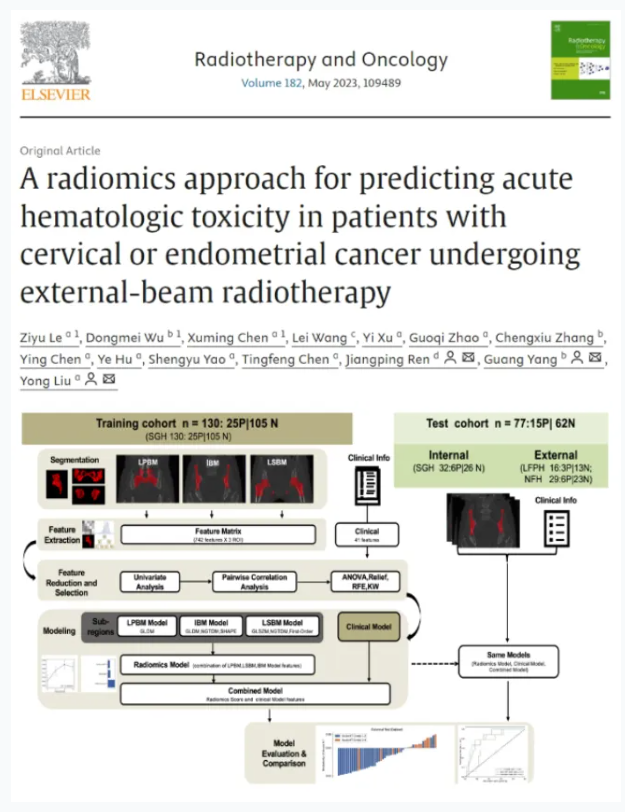
To tackle this issue, our team from Shanghai General Hospital collaborated with Professor Yang Guang’s team from East China Normal University. Together, they extracted radiomic features from pelvic images obtained through localization CT scans and combined them with other clinical characteristics to develop a composite predictive model. This model can forecast the risk of grade 3 or higher HT in patients. The results demonstrate that AI can assist in predicting and preventing HT caused by radiotherapy, with its effectiveness validated by multiple hospitals across China.
This achievement was published in 2023 in Radiotherapy and Oncology, the official journal of the European Society for Radiotherapy and Oncology (A radiomics approach for predicting acute hematologic toxicity in patients with cervical or endometrial cancer undergoing external-beam radiotherapy. Radiother Oncol. 2023;182:109489).